Parts Of The Eye / Anatomical Parts Of The Eye T Schlote Et Al Pocket Atlas Of Download Scientific Diagram : A thickened mass usually on the inner part of the white part of your eyeball.
Parts Of The Eye / Anatomical Parts Of The Eye T Schlote Et Al Pocket Atlas Of Download Scientific Diagram : A thickened mass usually on the inner part of the white part of your eyeball.. Without lubrication, the friction between the two layers of conjunctiva would cause rubbing. If you want to know what the human eye is like, enter here to find out. Parts of the eye (and what they do). It is disc shaped with a hole in the middle (the pupil). This intricacy is what makes eye anatomy such a fascinating subject.
The eye is a sensory organ. It allows you not only to view objects, but to see depth, color, size, and detail. Learn how eyes work by reviewing parts of the eye and their functions. Glaucoma is a result of the increased fluid pressure in the eye due to the reduction or blockage of aqueous from the anterior to posterior chambers. The eye itself is made of 10 general components that all work together to keep us seeing.
Parts Of The Eye Poster from knightowl.education Superior, inferior it is outermost supporting layer consists of thick membrane of tough fibrous connective tissue. This intricacy is what makes eye anatomy such a fascinating subject. Below, we discuss the different parts of the eye, as well as what issues can affect some of these components and therefore impact your ocular health. Protecting your eyes and the community. The eye is a sensory organ. This is especially important in the case of eye lasers, when a part of the cornea is removed in order to optimize the refractive power. We explain the parts of the eye in detail. It is the white color part of the eye which surrounds the cornea.
Parts of the eye and their functions.
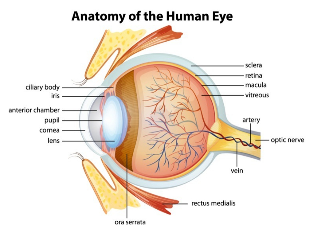
It is slightly thinner in the centre than the outer areas. Glaucoma is a result of the increased fluid pressure in the eye due to the reduction or blockage of aqueous from the anterior to posterior chambers. There are several physical and chemical elements that make up the eye. Near the front of the eye, in the area the most sensitive part of the retina is a small area called the macula, which has millions of tightly packed photoreceptors (the type called cones). The iris, which is the colored part of the eye, controls the amount of light that enters the eye. Parts of the eye outside the eyeball. We are a specialist ophthalmology center. Meet 10 eye parts that all work together to keep us seeing well every day. Additional parts of the orbicularis have been given separate names—namely, horner's muscle and the muscle of riolan; We explain the parts of the eye in detail. Aqueous humor generated from blood plasma. The eye has many parts which work together to accomplish vision, and to keep the structures required for vision safe from infection and injury. This intricacy is what makes eye anatomy such a fascinating subject.
Click on various parts of our human eye illustration for descriptions of the eye anatomy; If you know the parts of the eye, you will better understand how the eye works and how diseases and conditions can affect your vision. Light enters our pupil and is focused onto the retina at the back of the eye. They come into close relation opening of the eye is not just the result of passive relaxation of the orbicularis muscle but also is the effect of the contraction of the levator palpebrae. The iris is the coloured part of the eye (illustrated in blue above but in nature may be any of many shades of blue, green, brown, hazel, or grey).
Parts Of The Eye English Vocabulary Lesson Youtube from i.ytimg.com The sensitive surface of the eye needs to be kept moist. Read an article about how vision works. Superior, inferior it is outermost supporting layer consists of thick membrane of tough fibrous connective tissue. Learn how eyes work by reviewing parts of the eye and their functions. Eye anatomy, function, and physiology facts. The retina converts the light signal into electrical impulses. Light enters our pupil and is focused onto the retina at the back of the eye. Parts of the eye and their functions.
To enable clear vision, all structures within the eye should work effectively in order to capture light, focus it, and relay messages back to the brain to produce a visual image.
Parts of the eye (and what they do). See eye anatomy diagrams, definitions and more at lenscrafters. They come into close relation opening of the eye is not just the result of passive relaxation of the orbicularis muscle but also is the effect of the contraction of the levator palpebrae. The iris, which is the colored part of the eye, controls the amount of light that enters the eye. Parts of the eye, structure. The visible part of the eyeball makes up 1/6 of the eye's total surface area, with the rest hidden behind the eyelids. The eye is a complicated machine with many parts. We are a specialist ophthalmology center. Meet 10 eye parts that all work together to keep us seeing well every day. To enable clear vision, all structures within the eye should work effectively in order to capture light, focus it, and relay messages back to the brain to produce a visual image. The extraocular muscles are attached to the white part of the eye called the sclera. There are several physical and chemical elements that make up the eye. The outer or exterior shell is called sclera.
See eye anatomy diagrams, definitions and more at lenscrafters. Light enters the eye by passing through the transparent cornea and aqueous humor. The work of the visual system can be summarized as follows: The outer or exterior shell is called sclera. If you want to understand how various conditions and diseases affect the eye, then you need to know how the eyes work.
The Anatomy Of Your Eye Eyesite from www.eyesite.co.uk It is suspended from the ciliary muscles by the zonule. The eyes are in constant contact with your eyelids. Additional parts of the orbicularis have been given separate names—namely, horner's muscle and the muscle of riolan; To enable clear vision, all structures within the eye should work effectively in order to capture light, focus it, and relay messages back to the brain to produce a visual image. The sensitive surface of the eye needs to be kept moist. Parts of the eye (and what they do). The eye has many parts which work together to accomplish vision, and to keep the structures required for vision safe from infection and injury. The iris, which is the colored part of the eye, controls the amount of light that enters the eye.
Read an article about how vision works.
Additional parts of the orbicularis have been given separate names—namely, horner's muscle and the muscle of riolan; The work of the visual system can be summarized as follows: Learn how eyes work by reviewing parts of the eye and their functions. Read an article about how vision works. Parts of the eye and their functions. The conjunctiva also acts as a lining inside of your eyelid. It produces mucus and tears to lubricate your eyes and keeps microbes out of your eyes. The visible part of the eyeball makes up 1/6 of the eye's total surface area, with the rest hidden behind the eyelids. The eye sits in a protective bony socket called the orbit. In this post we'll examine all of the major parts of the eye, how they work, and what health conditions can affect them. The eye is also heavily involved with the nervous system, which allows the brain to take in information from the eyes and make the appropriate decisions on how to act upon this information. The eye is linked together with the nervous system, which allows the brain to take in information from the eyes and make the appropriate decisions on how to act upon this information. If you know the parts of the eye, you will better understand how the eye works and how diseases and conditions can affect your vision.
You have just read the article entitled Parts Of The Eye / Anatomical Parts Of The Eye T Schlote Et Al Pocket Atlas Of Download Scientific Diagram : A thickened mass usually on the inner part of the white part of your eyeball.. You can also bookmark this page with the URL : https://lilikuncoro.blogspot.com/2021/04/parts-of-eye-anatomical-parts-of-eye-t.html
Share Awesome
Belum ada Komentar untuk "Parts Of The Eye / Anatomical Parts Of The Eye T Schlote Et Al Pocket Atlas Of Download Scientific Diagram : A thickened mass usually on the inner part of the white part of your eyeball."


Belum ada Komentar untuk "Parts Of The Eye / Anatomical Parts Of The Eye T Schlote Et Al Pocket Atlas Of Download Scientific Diagram : A thickened mass usually on the inner part of the white part of your eyeball."
Posting Komentar